Virginia Equine Imaging
We're excited to share with our community that Virginia Equine Imaging has partnered with Asto CT to introduce cutting-edge Computed Tomography (CT) technology to our practice. VEI strives to offer compassionate care and individualized treatment to our community; with our new standing CT clients will receive a fast diagnosis through high quality images that can be acquired within 15 minutes.

Why Horse Owners Love the Asto CT Equina®:
Developed specifically for imaging both limbs simultaneously and the head & neck region in a mildly sedated standing equine patient, the Equina® is the world’s first dual-axis fan-beam computed tomography (CT) scanner. Your horse can maintain a natural standing position throughout the scan, eliminating the need for general anesthesia and reducing anxiety, ensuring a safe and more comfortable experience.

Unrivaled Precision
Computed Tomography (CT) is a noninvasive cross-sectional imaging modality that produces a 3D image of the scanned body part. It’s uses range from diagnosing bone injuries and complex fractures not visible on radiographs, screening horses for stress fracture pathologies before an athletic event, prepurchase exams, diagnosing soft tissue injuries such as tendon lesions, identifying thoracic or pelvic limb lameness, diagnosing diseases of the head, sinuses, teeth and jaw, surgical guidance, diagnosing neck issues such as osteoarthritis and more!
Case Studies
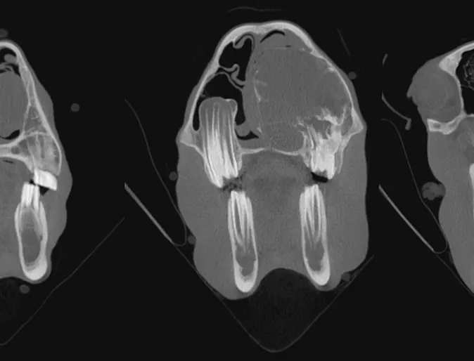
Aneurysmal Bone Cyst
9-month-old Percheron gelding presented for respiratory noise of 3 months duration. Final diagnosis on autopsy was Multilocular aneurysmal bone cyst.
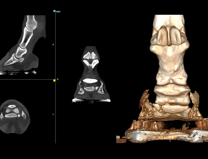
Fractured Navicular Bone
Percheron draft gelding with chronic right front limb lameness for over a year was diagnosed via Equine CT with a fracture in the lateral wing of the navicular bone, confirming the source of lameness.
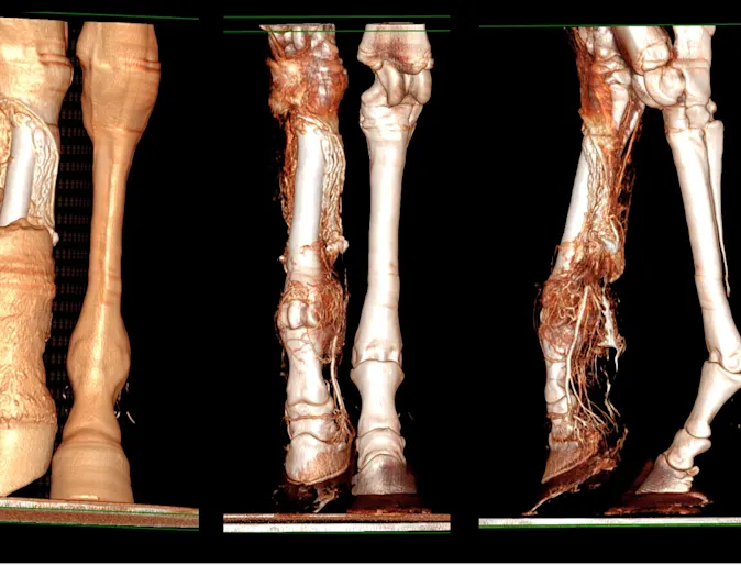
Distal Limb Perfusion
Yearling Warmblood filly suffered a degloving injury to the dorsal right hind metatarsus. A contrast CT was used to evaluate distal limb perfusion, guiding treatment to manage swelling and support healing.
Diagnostic Imaging Offered at VEI
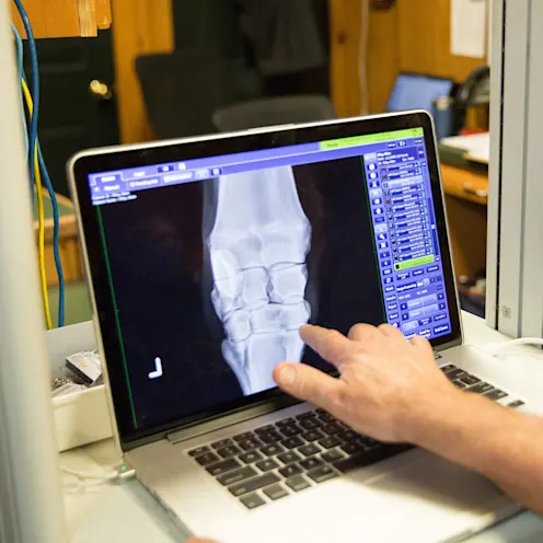
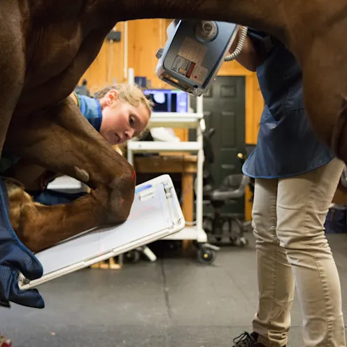
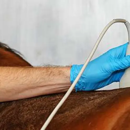
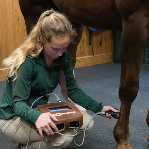
STANDING MRI - DIGITAL RADIOGRAPHY - DIGITAL ULTRASOUND- STANDING CT
